Fotografická soutěž
Katedra molekulární biologie a genetiky
Katedrová soutěž o nejlepší fotografii a obrázek
Soutěží se ve dvou kategoriích a v každé z nich je první cena 3000 Kč:
I. Kategorie "Vědecké zásluhy" - vystihuje zásadní průlom ve výzkumu z předchozího roku.
II. Kategorie "Estetika" - fotografie (obrázek) by měla být příjemná na pohled a otevřená všem druhům umělecké úpravy- lidé mohou uvolnit svého vnitřního surrealistu.
Soutěž je pod záštitou docenta Alexandera W. Bruce (
Soutěž vědeckých snímků Katedry molekulární biologie a genetiky pro rok 2023
(galerie fotografii z předešlých let zde)
Výsledky soutěže 2023
-
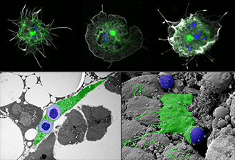
Gabriela Krejcova (Adam Bajgar lab)
SCIENTIFIC MERIT PRIZE
1st place
GPs: Macrophage-specific delivery tool visualized three ways. Confocal and electron microscopy images of Drosophila macrophages (green) internalizing yeast-derived glucan particles, which represent a suitable macrophage-specific delivery tool.
-
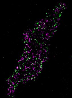
Alexey Bondar (Hana Sehadova lab)
SCIENTIFIC MERIT PRIZE
2nd place
Single molecule interactions - the image demonstrates interactions between individual signaling molecules (a G protein-coupled receptor and a G protein) in living cells after treatment with dopamine.
-
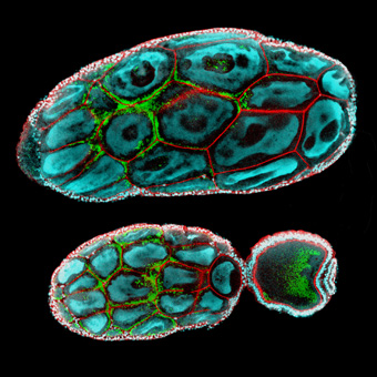
Hana Sehadova (Sehadova lab)
SCIENTIFIC MERIT PRIZE
3rd place
Rocket to Mars - Parts of the queen bumblebee's ovary. The developing oocyte is nourished by a group of sister cells.
-
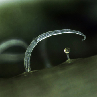
Hana Sehadova with Oxana Skokova-Habustova (Sehadova lab)
AESTHETIC MERIT PRIZE
1st place
Inseparable friends - A muscular and glandular trichome on a potato leaf of the Marabel variety.
-
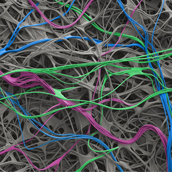
Hana Sehadova with Michal Zurovec (Sehadova lab)
AESTHETIC MERIT PRIZE
2nd place
A tangled path - The silk of the spindle ermine moth, Yponomeuta cagnagella.
-
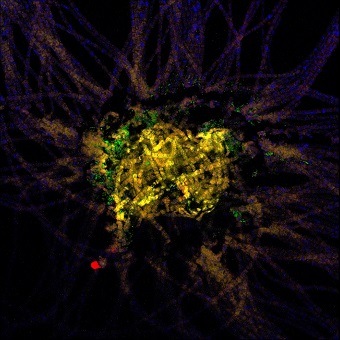
Anxhela Hania (Sobotka lab)
AESTHETIC MERIT PRIZE
3rd place
Colony core of Trichodesmium - a marine filamentous cyanobacterium; the sample was collected in the Gulf of Aqaba (Eilat, Israel) and employed to visualize the presence of associated bacteria.
-
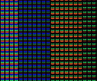
Thinles Chondol (Jiri Dolezal lab)
Cross-section of the root of plant Askellia flexusa (Asteraceae) - from the Himalayas (~ 4700 m asl) stained with AstraBlue and Safranin solution (Abbreviations: ep - epidermis; ph - phloem; xv - xylem vessel; xf - xylem fiber; pa - parenchyma; ca - cambium; mr - medullary rays).
-
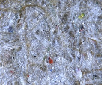
Alexey Bondar (Hana Sehadova lab)
Recycled paper - the photo of a paper towel made from recycled paper with visible bits of different candy wrappers from the original paper used for recycling.
-
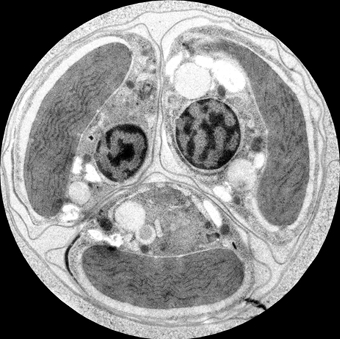
Shu-Min Yang (Miroslav Obornik lab)
An ultrathin TEM section of a four-cell cyst of Chromera velia
-
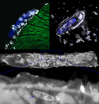
Gabriela Krejcova (Adam Bajgar lab)
Wasp egg: Immune response to a parasitoid egg. Although the parasitoid wasp egg (blue) laid in Drosophila larva (visualized by micro computed tomography) tries to hide from the host immune response in the folds of intestine (green), it is recognized by plasmatocytes (white, upper left) and later encapsulated by lamellocytes (white, upper right).
-
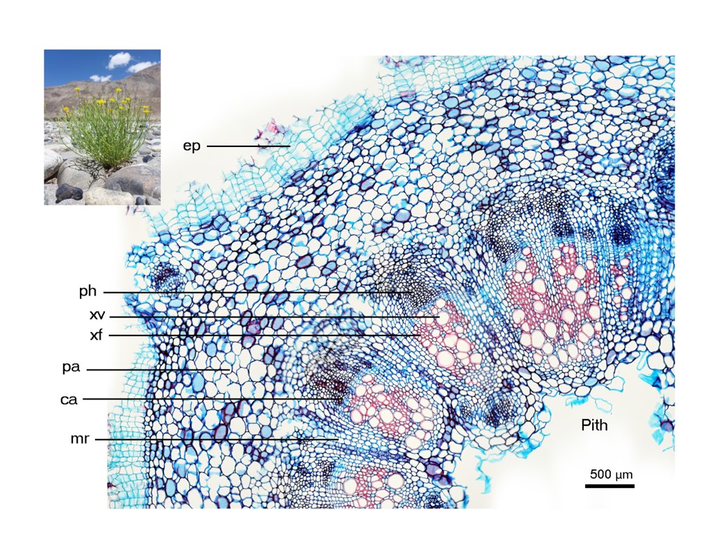
Thinles Chondol (Jiri Dolezal lab)
Cross-section of the root of plant Askellia flexusa (Asteraceae) - from the Himalayas (~ 4700 m asl) stained with AstraBlue and Safranin solution (Abbreviations: ep - epidermis; ph - phloem; xv - xylem vessel; xf - xylem fiber; pa - parenchyma; ca - cambium; mr - medullary rays).
-
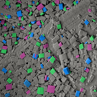
Hana Sehadova with Ivo Sauman (Sehadova lab)
Crystals - Calcium oxalate crystals on the silk cocoon of the cecropia moth, Hyalophora cecropia.
-
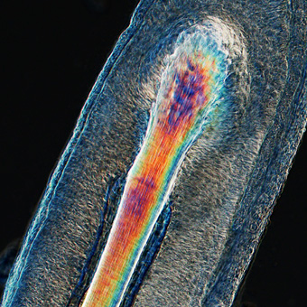
Hana Sehadova (Sehadova lab)
On the hair fiber - Detail of a hair root of a curious student. The image was taken during a practical microscopy exercise.
-
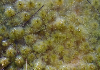
Zuzana Urbanova (Urbanova lab)
Frozen beauty of Sphagnum cuspidatu.
Archiv fotografii 2011-2022
- Přečteno: 8365
