Selection of microscopic images
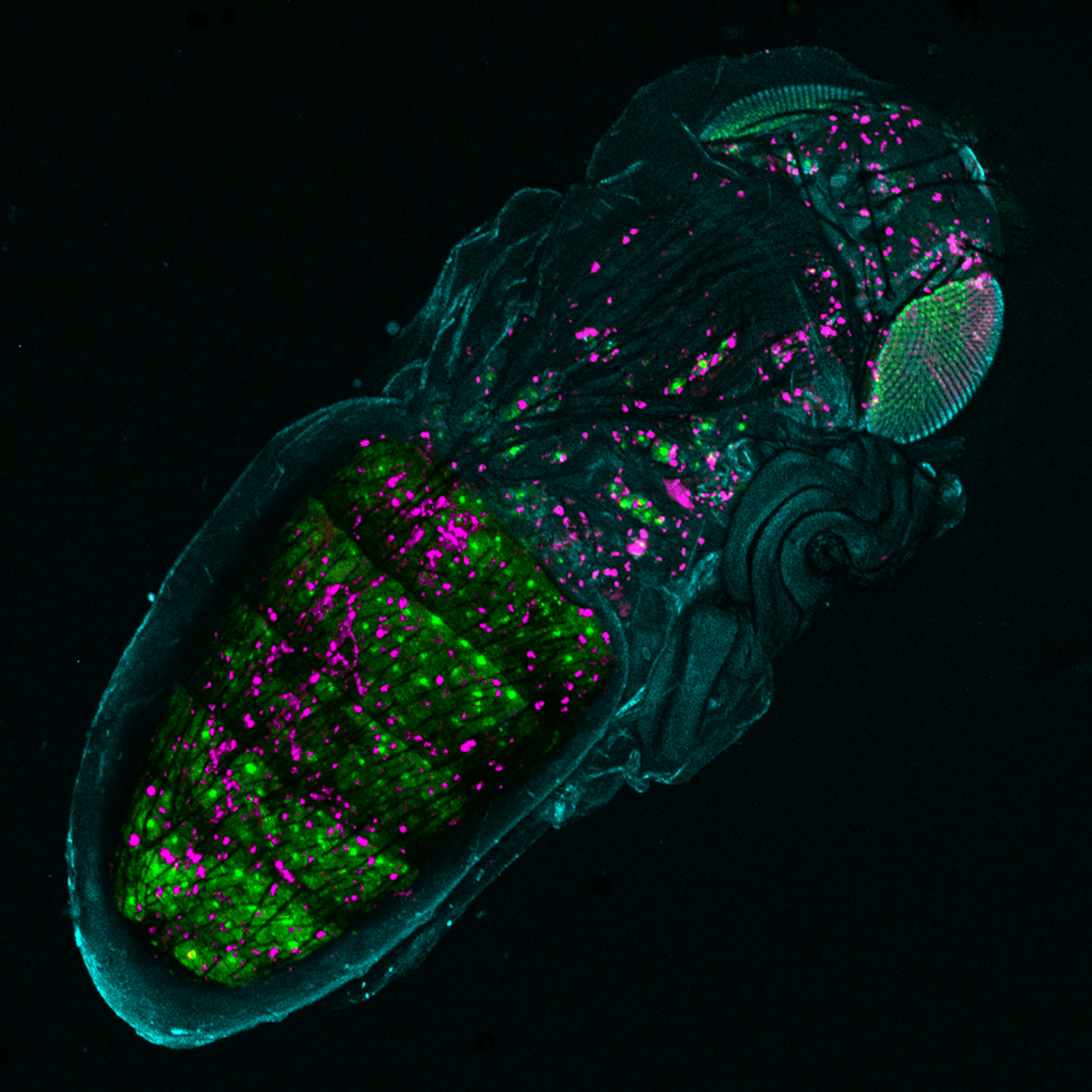
Image 1: Confocal microscopy image of Drosophila melanogaster pupa depicting interaction of immune cells (magenta) with histolyzing larval adipocytes (green). Image captured by the members the of Laboratory of Insect Metabolism, Faculty of Science, University of South Bohemia, Czech Republic.
Author: Mgr. Gabriela Krejčová (
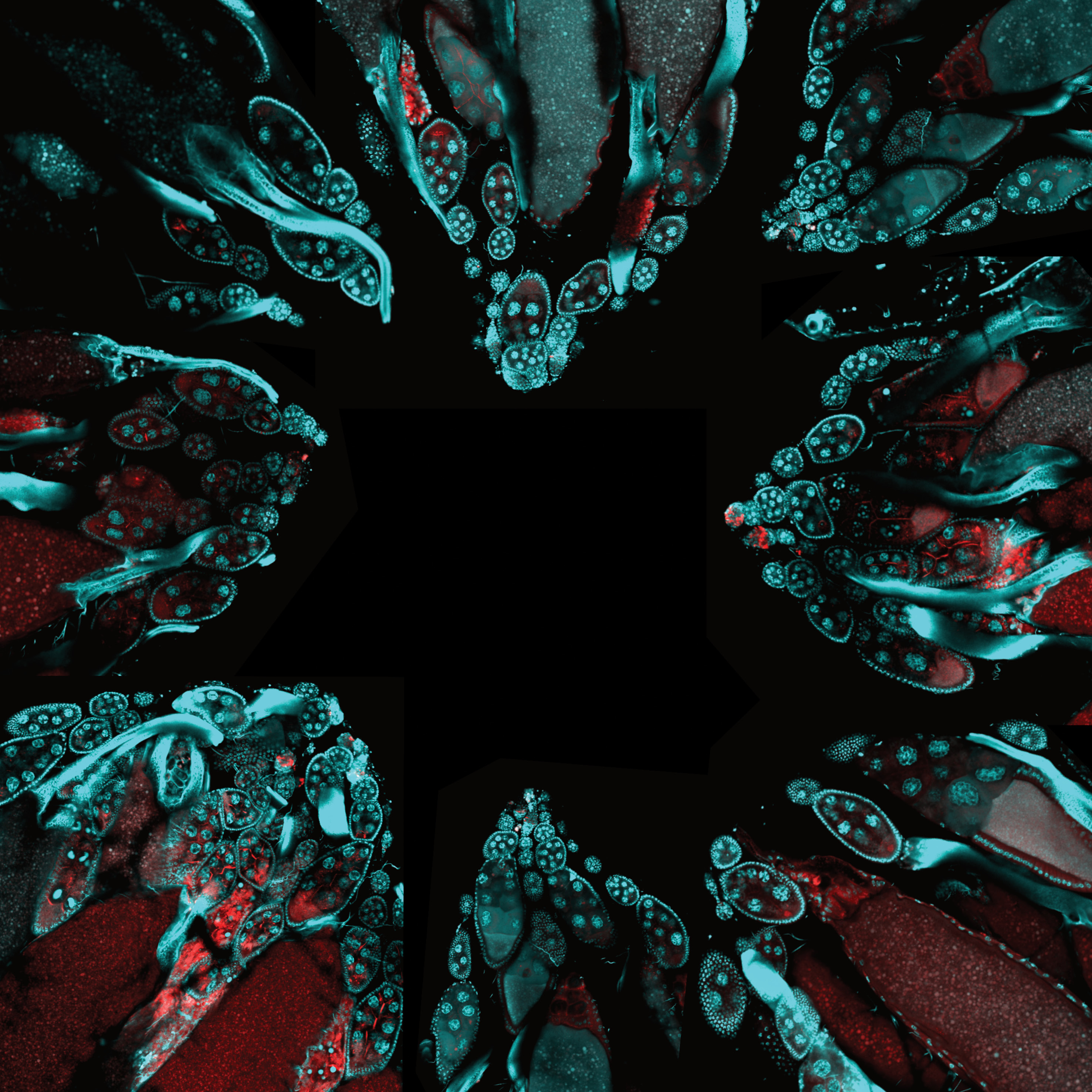
Image 2: Composition created from confocal images of dissected ovaries of Drosophila melanogaster. Nuclei stained by DAPI (cyan); membranes marked by phalloidin (red). Image captured by the members the of Laboratory of Insect Metabolism, Faculty of Science, University of South Bohemia. Image captured by the members the of Laboratory of Insect Metabolism, Faculty of Science, University of South Bohemia, Czech Republic.
Author: Mgr. Gabriela Krejčová (
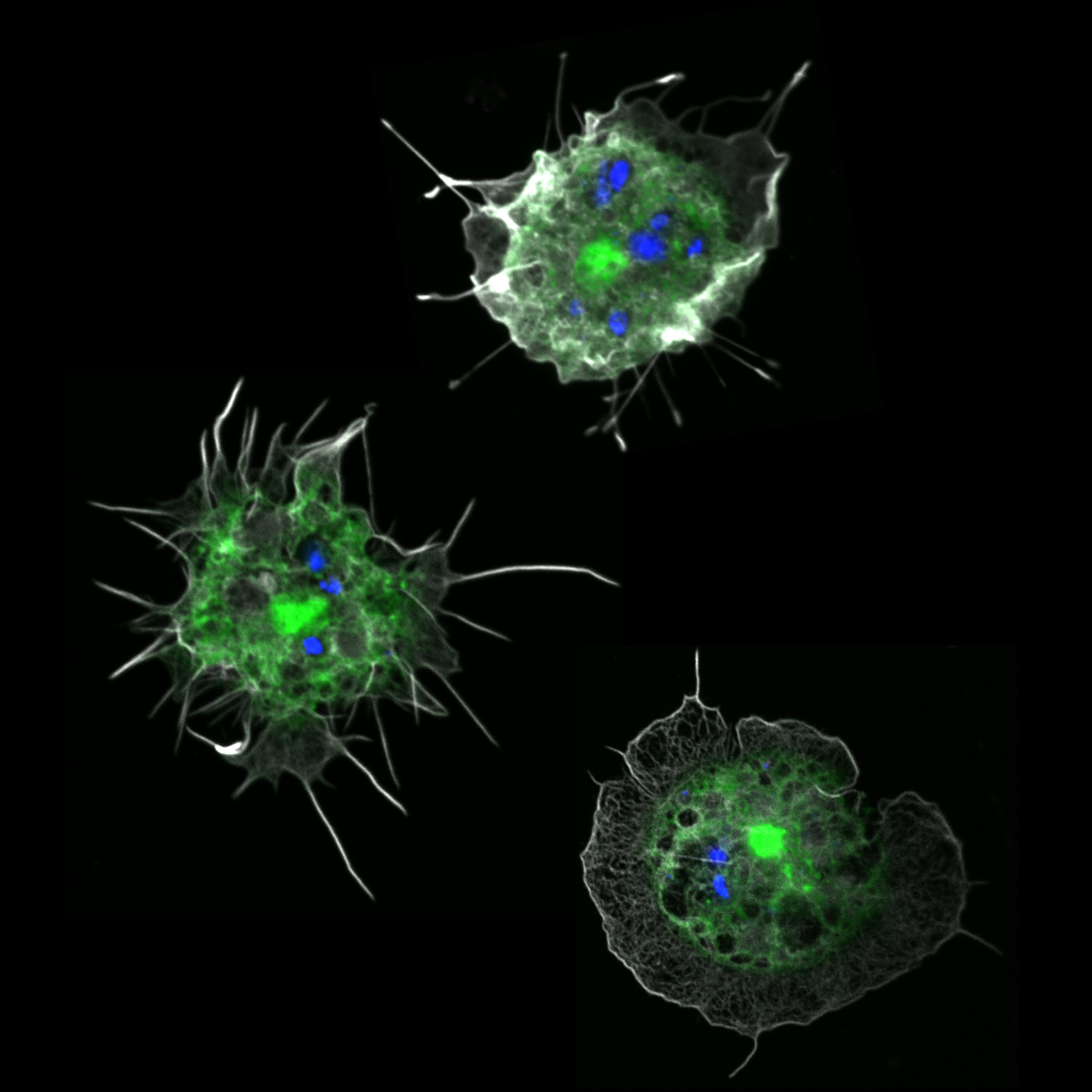
Image 3: Confocal images of macrophages of Drosophila melanogaster, which contain glucan particles suitable for macrophage specific-delivery of various metabolic inhibitors or transcription factors developed in the Laboratory of Insect Immunometabolism (Faculty of Science, University of South Bohemia). Macrophages were visualized by endogenously produced GFP under croquemort driver. Blue color marks glucan particles; white color depicts macrophage membrane. Image captured by the members the of Laboratory of Insect Metabolism, Faculty of Science, University of South Bohemia, Czech Republic.
Author: Mgr. Gabriela Krejčová (
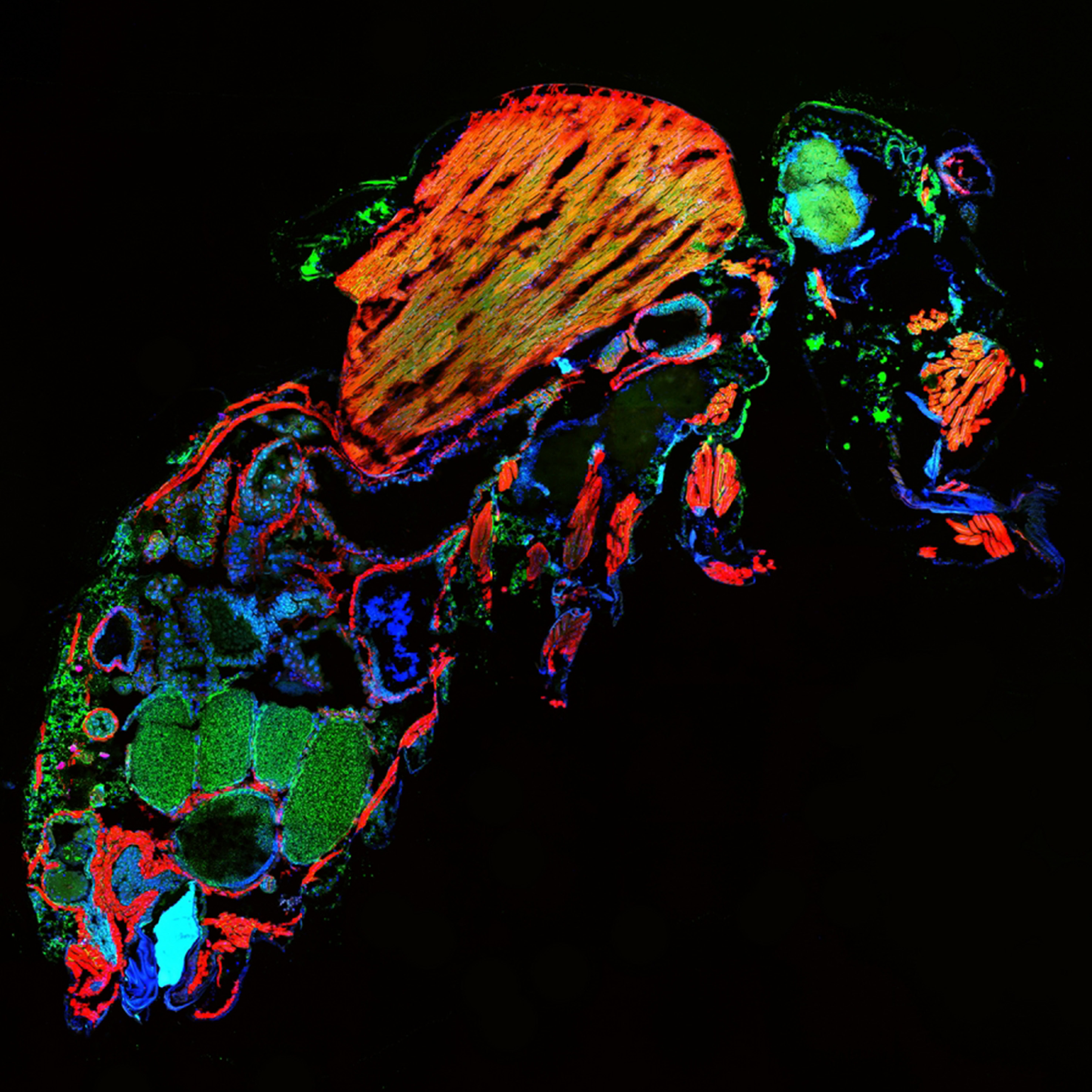
Image 4: Histological cross-section of Drosophila melanogaster female. Actin was stained by phalloidin (red), nuclei by DAPI (blue), and lipids by BODIPY (green). Image captured by the members the of Laboratory of Insect Metabolism, Faculty of Science, University of South Bohemia, Czech Republic.
Author: Mgr. Gabriela Krejčová (
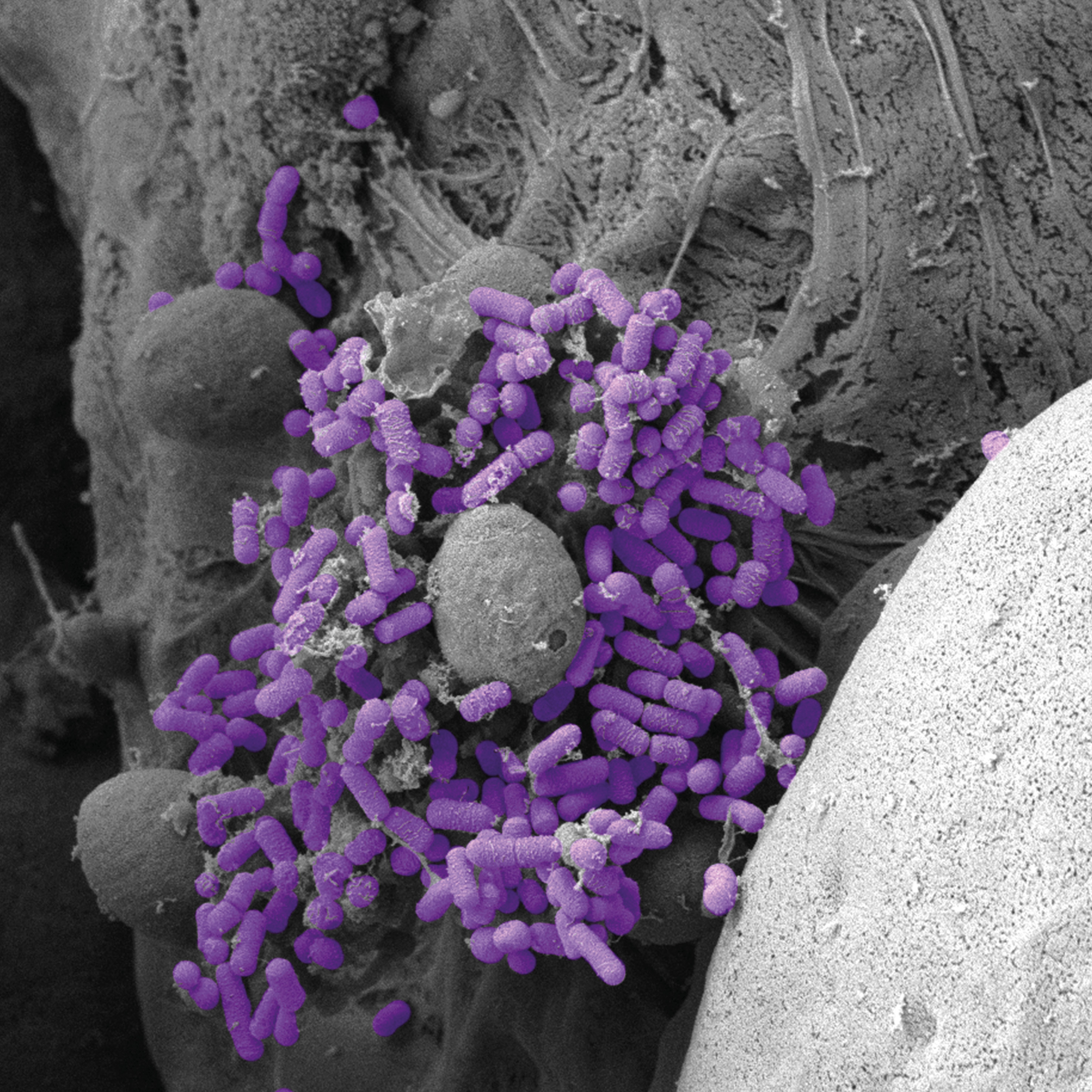
Image 5: Scanning electron microscopy image of Drosophila melanogaster macrophage with attached bacteria (Streptococcus pneumoniae; in violet). Image captured by the members the of Laboratory of Insect Metabolism, Faculty of Science, University of South Bohemia, Czech Republic.
Author: Mgr. Gabriela Krejčová (
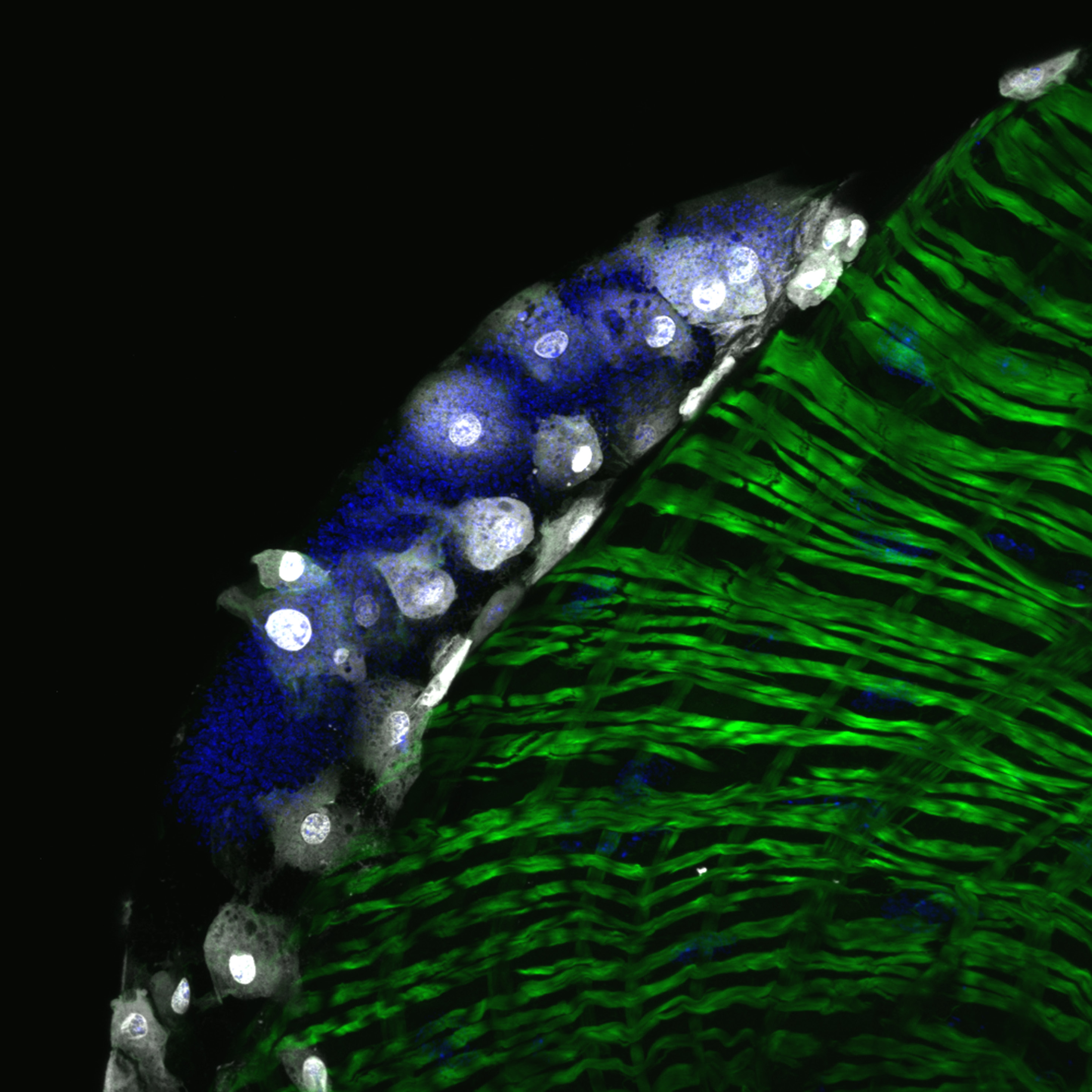
Image 6: Confocal image of an egg laid by the parazitoid wasp Leptopillina boulardi in the body cavity of Drosophila melanogaster larva. Although it tries to hide in the folds of the intestine (gut musculature in green), it is recognized by immune cells (white). Image captured by the members the of Laboratory of Insect Metabolism, Faculty of Science, University of South Bohemia, Czech Republic.
Author: Mgr. Gabriela Krejčová (
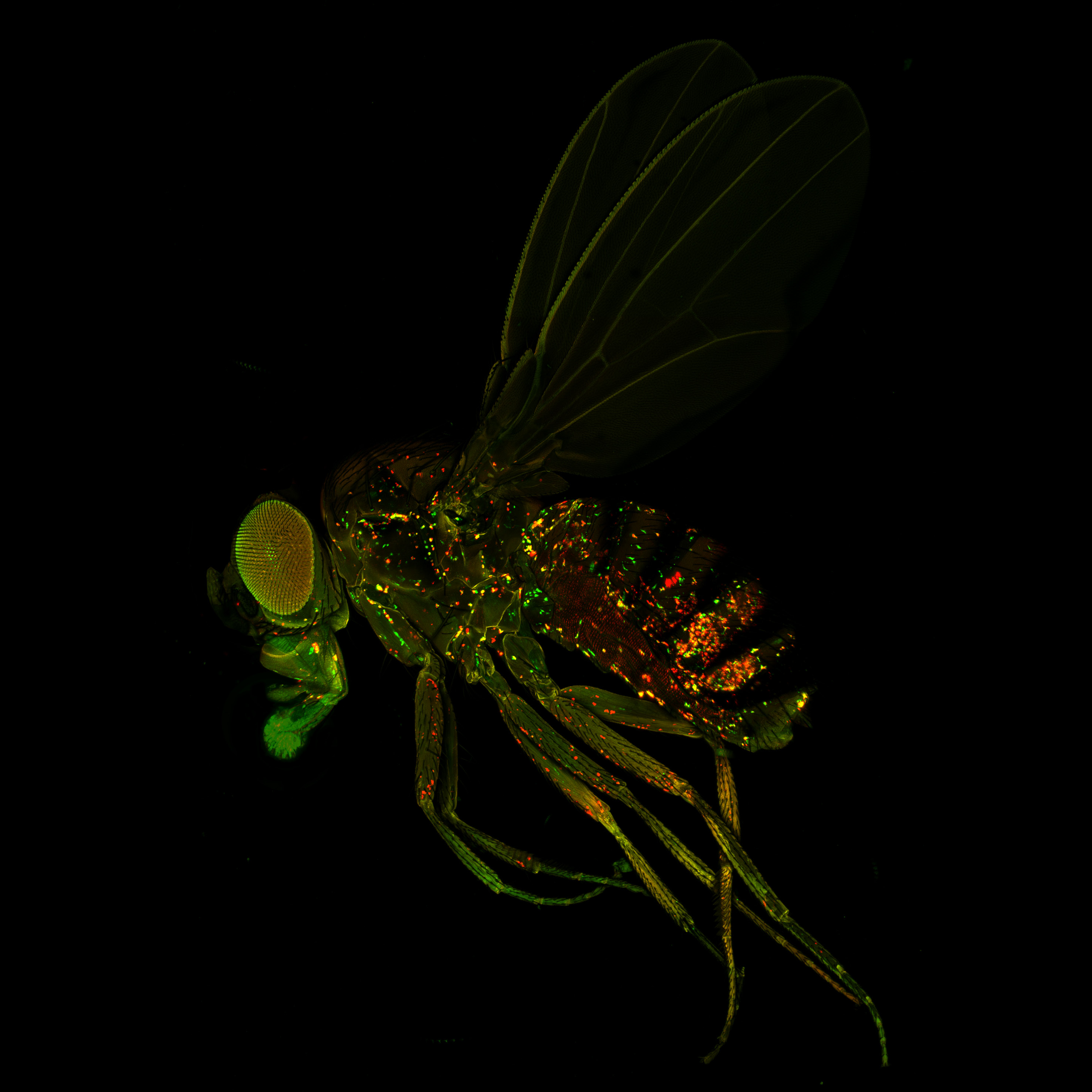
Image 7: Confocal image of Drosophila melanogaster showing increased expression of lactate dehydrogenase (in red) in immune cells (in green) after bacterial infection. Image captured by the members the of Laboratory of Insect Metabolism, Faculty of Science, University of South Bohemia, Czech Republic.
Author: Mgr. Gabriela Krejčová (
- Hits: 247